
When Professor Panicos Kyriacou received a visit from a team of neurosurgeons from the Royal London Hospital asking for his help, their meeting led to a ground-breaking research project that is set to transform the monitoring and treatment of patients suffering from traumatic brain injuries (TBI). TBI is the most common cause of death and disability in the under 40 age group both in the UK and worldwide and prevalence is increasing.
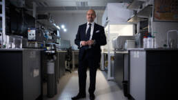
Professor Kyriacou is Director of the Research Centre for Biomedical Engineering (RCBE) at City, University of London, which has gained a global reputation for its pioneering work in developing complex medical devices, optical biosensors and signal and image processing techniques to diagnose and treat the sick and injured.
His personal research interest focuses primarily on non-invasive optical sensors to detect biomarkers relating to disease and this was the reason for the visit by the neurosurgeons.
“They came to our lab and asked whether it was possible to measure or monitor intracranial pressure (ICP) in traumatic brain injury patients non-invasively,” Professor Kyriacou explained.
“ICP is defined as the pressure within the skull and brain. TBI often causes a rise in ICP as the brain swells within the rigid skull and therapy is directed at keeping this pressure at an acceptable level with medications or surgery. Very high ICP may lead to further brain damage resulting in increased disability or death. Existing gold standard techniques to measure ICP and cerebral oxygenation involve placing a sensor into the brain tissue through a small hole drilled in the skull. This invasive procedure risks infection and bleeding into the brain and can only be performed by a neurosurgeon. Therefore, there is a vital demand to develop non-invasive technologies that will allow continuous measurements of ICP.”
As a first approach to the problem, Professor Kyriacou and colleagues from the RCBE made a trip to the neurocritical care unit at the Royal London to observe how surgeons monitored patients with serious traumatic brain injuries using existing technology.
He said: “I watched the surgeons drill a hole in the skull of a patient and then they inserted a pressure transducer. They literally stabbed this sensor right into the brain. It must be the most invasive, non-therapeutic procedure.”
Harnessing the knowledge built up by 25 years of research into how light behaves with tissue or blood, Professor Kyriacou and his team set about developing the world’s first non-invasive optical sensor to monitor intracranial pressure.
“We knew very well that we can send light through the forehead, into the skull and into the brain,” he said. “Light it comes in different colours –visible colours, near, mid and far infrared – and we knew from studies we had done in our lab that certain colours or certain wavelengths of light can penetrate into the brain.”
Developing the sensor was made possible by the combination of advanced medical optics and advanced computational techniques, drawing upon the knowledge of experts in computer science, machine learning and artificial intelligence.
“The sensor is placed on the forehead and we shine light into the brain through the skull,” explained Professor Kyriacou. “The light bounces around, but some light is reflected back. We harness the light coming back – at different wavelengths – and then we apply machine learning models to tell us how that light is affected by hemodynamic changes and intracranial pressure in the brain.
“When the pressure is rising in the brain, the vessels in the brain are compressed. This rising pressure has an impact on the flow of blood and the viscoelastic properties of the arteries. These changes are manifested on the optical signals, also called Photoplethysmograms (PPGs), we acquired from the brain. Utilising advanced computational models on the acquired optical signals enables the prediction the intracranial pressure.”
The sensor was developed through in vitro control experiments in the lab, using ‘phantoms’ of the brain to simulate what happens when a patient suffers a traumatic brain injury. Once the concept was proved, it went to clinical trials before receiving formal approval from the UK Government’s Medicines and Healthcare Products Regulatory Agency.
The next step was to introduce the sensor to neurocritical care to assess its performance against the traditional invasive method of monitoring pressure within the brain. Some 50 traumatic brain injury patients being treated with the invasive probe in situ also had the non-invasive optical sensor attached to their forehead and the results were compared.
“We collected the data and the estimation of intracranial pressure through our optical sensors was too good to be too true,” said Professor Kyriacou. “It was amazing. The correlations were extremely high.”
The success of the project means the optical sensor could soon be in widespread use neurocritical units. With the support of the RCBE team and clinicians, the University has secured the intellectual property of the device and has recently licensed the technology to a consortium in the Netherlands, who plan to establish a UK-based company to take it forward to commercialisation.
As a Professor of Biomedical Engineering, the journey from basic research to applied research and then to commercial application – from the lab to the bedside – is hugely rewarding. Professor Kyriacou studied engineering as an undergraduate but his passion for medicine led him to dedicate his research career to developing technology solutions for healthcare.
“As a biomedical engineer, this is my vision,” he said. “I enjoy doing research but what I enjoy more is to be able to see the translation of these technologies to the bedside to benefit patients to improve the quality of life to patients, their carers and their families.”
The intracranial pressure monitor is just one of numerous innovations developed by Professor Kyriacou and his colleagues, that is pushing the boundaries of optical sensor technologies.
Most of this work is applied to physical conditions such as cardiovascular, neurodegenerative or blood diseases, but the research effort has been expanded to include mental health, specifically for people suffering from bipolar disease.
Bipolar patients are generally prescribed lithium to prevent them swinging between manic and depressive moods, but the dosage has to be precise. Take too much and it can cause irreversible damage to kidneys; take too little and it fails to stabilise the mood swings.
The problem for patients is confirming that their lithium is at the right level. This has to be done by providing regular blood samples at their GP surgery, which discourages many patients from taking the drug, while many fail to turn up to their appointments. Even for those who take it diligently and attend their appointments, results are not available until their sample has been sent to a lab, analysed and then reported to the GP.
To address this issue, Professor Kyriacou and fellow researchers conducted experiments in the lab to prove that lithium has a distinct optical signature, enabling them to develop optical sensor technology to detect lithium concentrations in the blood.
They have now reached the stage of testing the sensor on patients – either on the skin or by taking a droplet of blood – with the aim of producing either absolute measurements of lithium levels or otherwise a traffic-light warning system indicating whether a patient should consult their GP.
If successful, the technology would be transformative by allowing bipolar patients to test themselves at home, providing reassurance that their lithium level is in the therapeutic range or that they need to seek advice.
As part of their activities in designing, fabricating and evaluation optical technologies, Professor Kyriacou and his team work closely with London hospitals such as St Bartholomew’s, the Royal London and Great Ormond Street Hospital for Children.
The latter has engaged Professor Kyriacou with numerous challenges over the years, one of which was the question of whether it was possible to use sensor technology to monitor oxygenation in the brain of a new-born baby to check for hypoxic encephalitis – a pathology that means not enough oxygen is going to the brain, with all the severe consequences that brings. The alternative is to carry out an MRI scan, but this is expensive and resource-heavy and does not facilitate continuous, dynamic monitoring.
Professor Kyriacou says the soft spot on a new-born’s forehead – the anterior fontanelle – offers the perfect optical window to shine light into the baby’s brain and using wavelengths responsible for oxygenation, they were able to develop the necessary sensors to calculate oxygen levels.
He said: “I remember vividly when we did the study, nurses from intensive care, clinicians and anaesthesiologists were gathered round and were saying, ‘Oh my goodness, are you really interrogating the brain with these sensors?’ The signals were amazing and the calculations for oxygenation were very good, even for a prototype device.”
Interest in the area of non-invasive optical technologies has grown exponentially over the last two decades, according to Professor Kyriacou and he can only see it getting bigger, whether it is medical devices for use in clinical practice or wearable devices to monitor wellbeing.
His hope is that the growth of wearables will make a global contribution to saving lives in low-resource countries by a providing a means to monitor vital signs as a way of screening against the onset of an infection.
“The mortality of children under five around the world is staggering,” he said. “You cannot comprehend the numbers. You would have thought it would be impossible in the 21st century for so many children under five to die because they cannot diagnose a simple infection.
“At the end of the day, we are in the business of creating healthcare solutions for everybody, not selective nations or countries.
“For me, creating technologies where they can contribute globally in low-resource environments and save the lives of children because they don’t have basic technology – that is fundamental to the work we do in the lab.”